Introduction
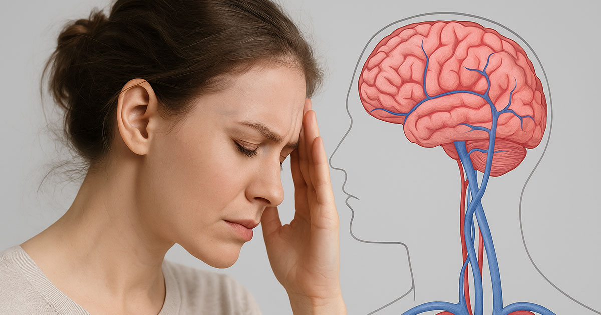
In recent years, much attention has been given to cerebral venous drainage and its impact on neurological health. The idea that impaired venous outflow from the brain can disrupt intracranial balance—triggering disabling symptoms such as headaches, visual disturbances, or cognitive deficits—has attracted growing interest, especially following the description of CCSVI (Chronic Cerebrospinal Venous Insufficiency).
At the same time, the scientific community has begun to look with fresh eyes at conditions that are considered rare—though perhaps not as rare as once thought: connective tissue disorders, particularly Ehlers-Danlos syndrome (EDS). Those living with EDS are all too familiar with the widespread fragility it brings: unstable joints, chronic pain, vascular problems, and neurological symptoms that are often difficult to explain through standard imaging alone.
Two recent publications – one in Frontiers in Neurology, and another reported by Ehlers-Danlos News – have shed light on a clinical picture that had long remained in the shadows. When a patient with EDS also presents with cerebral venous drainage disorders, we are not simply faced with the sum of two diagnoses. Instead, what emerges is a new clinical phenotype, with its own characteristics: more severe, more complex, and carrying specific diagnostic and therapeutic implications.
This seemingly subtle observation is in fact a key turning point: recognizing a new phenotype means acknowledging that there is a category of patients who cannot be treated with the same protocols as others. It means that physicians and researchers must pause, listen, and study more carefully a phenomenon that challenges the usual tendency to oversimplify diagnoses.
In the article that follows, we will take a closer look at this connection: what EDS is and why it predisposes patients to worsening cerebral venous drainage, what the latest studies reveal, and what practical consequences this has for patients’ quality of life. We will do so in a clear and conversational style, without compromising scientific accuracy, because the goal is simple: to help those living with these conditions feel seen, and to encourage those treating them to look with fresh eyes.
Ehlers-Danlos and Cerebral Venous Drainage
Ehlers-Danlos Syndrome (EDS) is not a single disease, but rather a group of more than ten genetic variants, all linked by a defect in connective tissue. In practical terms, the “biological glue” that holds together organs, joints, blood vessels, and skin is faulty. The consequences include hypermobile joints, fragile skin, abnormal scarring, and—in the most severe cases—vascular rupture.
Traditionally, EDS has been thought of as an “orthopedic” condition, tied to joints that dislocate easily or to chronic musculoskeletal pain. But connective tissue is not limited to the joints—it is found throughout the entire body. This means that EDS is a systemic disorder, with manifestations that also involve the neurological and vascular systems.
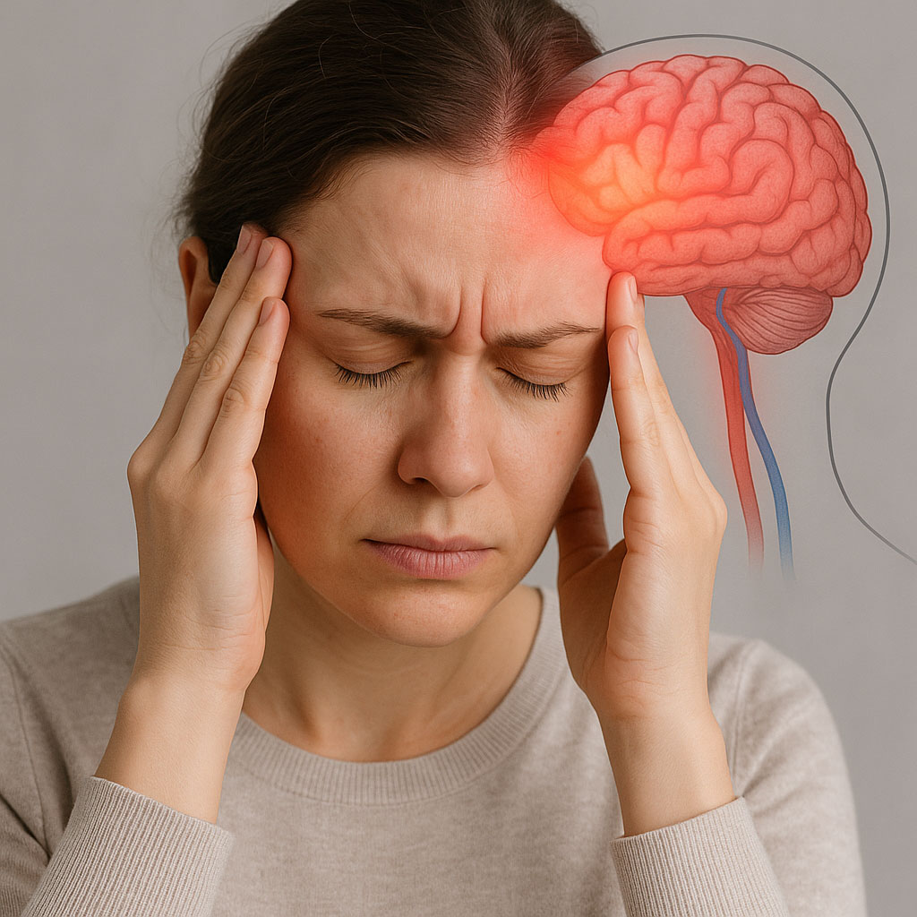
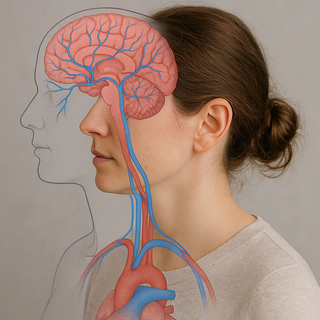
And this is where cerebral venous drainage comes into play. Like every other organ, the brain requires a constant blood flow: arteries to deliver oxygen and nutrients, and veins to drain carbon dioxide and metabolic waste. When venous outflow is obstructed—by stenosis, compression, or collapse—intracranial pressure is disrupted, tissues become hypoxic, and waste products and toxic proteins begin to accumulate.
In patients with EDS, however, fragile connective tissue does not adequately support venous structures. This increases the risk of veins collapsing or dilating abnormally, compromising proper drainage. It is as if the framework meant to keep the vessels open is too weak to withstand mechanical and pressure-related stress.
The resulting symptoms are not always dramatic, but they are often chronic and debilitating:
- Persistent headaches, which worsen when standing upright.
- Visual disturbances, such as blurred vision, double vision, or a sensation of ocular pressure.
- Brain fog, with difficulty concentrating and slowed cognition.
- Postural instability and dizziness, resembling idiopathic intracranial hypertension.
Postural instability and dizziness, resembling idiopathic intracranial hypertension.
These symptoms are so common among EDS patients that they are often dismissed as “nonspecific” or “functional.” Yet new research shows they in fact have a clear organic basis: impaired venous drainage.
In other words, EDS not only predisposes patients to orthopedic and general vascular problems, but also creates the perfect conditions for worsening cerebral venous disorders. And this is where the concept of a distinct clinical phenotype emerges.
The New Clinical Phenotype
The study published in Frontiers in Neurology took a retrospective approach, analyzing cases of patients with connective tissue disorders and cerebral venous drainage abnormalities. What emerged was not simply an accumulation of diagnoses, but a recurring pattern—a constellation of features appearing with striking consistency.
The data speak for themselves:
- 87% of patients were female.
- The average age was 36, confirming that this is not an issue of the elderly but of young adults, often in the midst of their working and family lives.
- Symptoms were more severe than in patients with CVD alone, with more intense headaches, greater disability, and marked cognitive impairment.
- Quality of life was significantly lower, with daily limitations far beyond the average.
- Adverse drug reactions and surgical complications were frequent, due to connective tissue fragility and increased sensitivity to materials and anesthetics.
From these observations arises the concept of a new clinical phenotype: patients with EDS and cerebral venous disorders who share common features significant enough to represent a distinct entity.
Why does this matter? For at least three reasons:
- Diagnosis – A patient with EDS who presents with chronic headaches and visual disturbances should not be dismissed as a “somatizer” or given a vague label such as FND. There is a concrete possibility that this is a cerebral venous disorder aggravated by EDS.
- Therapy – Treatment strategies that may be safe and effective for patients without EDS are not automatically suitable for those with connective tissue fragility. What is needed is a personalized approach that avoids unnecessary risks.
- Research – Recognizing a new phenotype paves the way for dedicated studies, targeted protocols, and, ultimately, specific guidelines.
This step is crucial because it changes the physician’s perspective: no longer a “difficult patient” or a “complicated case,” but rather a well-defined clinical profile that deserves particular attention.
The very concept of a clinical phenotype carries significant weight in medicine. It means that a recurring set of symptoms and signs observed in multiple patients has scientific validity and can be studied as a distinct entity. In the case of EDS associated with cerebral venous disorders, this recognition delivers a clear message: we are not dealing with coincidences, but with a specific pathological mechanism.
Practical Implications
Understanding that a new clinical phenotype exists—the coexistence of EDS and cerebral venous drainage disorders—is not just an academic exercise. The implications are very concrete and touch on three central areas: symptoms, diagnosis, and treatment.
Typical and aggravated symptoms
Cerebral venous disorders in EDS patients are not only more frequent but often more severe. Many report daily headaches that worsen when standing and tend to improve when lying down or during cervical traction. This is not the classic migraine: it is a pressure-like pain, sometimes accompanied by nausea, dizziness, and double vision.
Other reported symptoms include:
- Blurred or double vision, linked to increased intracranial pressure compressing the optic nerves.
- Cognitive disturbances (“brain fog”), manifesting as difficulty concentrating, slowed thinking, and short-term memory loss.
- Postural instability, with dizziness and sudden falls.
- Otological symptoms, such as tinnitus and a sensation of ear pressure, often worsening in an upright position.

Many of these symptoms are nonspecific and can be mistaken for anxiety, depression, or psychosomatic conditions. It is precisely this ambiguity that leaves many EDS patients wandering for years without an accurate diagnosis. The Frontiers study, however, demonstrates that there is a clear organic basis: altered venous drainage amplified by connective tissue fragility.
Differential diagnosis: a necessary challenge
Diagnosis in these cases is far from straightforward. Standard radiological studies often fail to detect the subtler abnormalities, especially the dynamic ones that change with posture. A brain MRI performed in the supine position may appear normal, while the patient continues to report severe symptoms.
A more sophisticated diagnostic approach is therefore required:
- Doppler ultrasound of the jugular and vertebral veins, ideally performed both upright and supine to observe postural variations.
- Venography with pressure measurements, useful in complex cases to evaluate stenosis or reflux.
- Dynamic venous imaging, such as MR venography with specific protocols.
- Comprehensive clinical assessment, taking into account the patient’s EDS history, allergies, and vascular fragility.
The key point is not to stop at the first “normal” image. In a patient with EDS and neurological symptoms, the suspicion of a venous disorder must remain alive, even if initial tests fail to show obvious abnormalities.
Personalized therapy
Treatment may represent the greatest challenge. Strategies that work for patients without EDS are not automatically transferable.
- Pharmacology – Some medications used to reduce intracranial pressure (such as acetazolamide) can provide benefits, but they require close monitoring due to potential side effects. Moreover, EDS patients often experience more frequent adverse reactions or intolerances.
- Surgery – Procedures such as venous angioplasty must be evaluated with extreme caution, given the connective tissue fragility that increases the risk of complications.
- Postural and conservative approach – Sleeping with the torso elevated at a 30–35 degree angle, avoiding positions that compress the neck, and using collars or cervical supports during certain activities can help improve symptoms.
- Multidisciplinary follow-up – Neurologists, geneticists, phlebologists, neurosurgeons, and physiotherapists must work together, as no single specialist can manage the complexity of these cases alone.
The real lesson from this new phenotype is clear: there is no universal protocol. Every patient must be evaluated individually, with an approach that considers their fragility and specific clinical manifestations.
CCSVI and Clinical Analogies
The description of the new EDS + CVD phenotype fits into a broader context: that of CCSVI. Over years of clinical practice, CCSVI has been described as a condition characterized by stenosis, reflux, and compression of the extracranial veins, which disrupt the brain’s hemodynamic balance.
The symptoms of CCSVI often overlap with those reported in the study on EDS and venous drainage:
- Chronic headaches.
- Visual disturbances.
- Cognitive symptoms and mental fatigue.
- Tinnitus and dizziness.
In both cases, posture plays a crucial role. Many patients report that their symptoms worsen when upright and improve when lying down or with cervical traction—an indication of a dynamic, rather than fixed, condition.

What distinguishes the EDS phenotype is its underlying structural fragility: while in classic CCSVI stenosis and reflux are often linked to external compressions (muscles, bones, anatomical anomalies), in EDS patients the weakness of the connective tissue makes the veins inherently unstable. This aggravating factor leads to a more severe clinical presentation.
We can therefore say that:
- CCSVI provides a useful paradigm for interpreting cerebral drainage disorders.
- The EDS + CVD phenotype is a particular variant, with its own specific features that must be recognized.
This analogy is not purely theoretical. It has direct implications for both research and clinical practice. The tools and methodologies developed in the study of CCSVI can be adapted and applied to better understand the EDS phenotype—provided that the greater fragility of these patients is carefully taken into account.
A concrete example is sleep management in an inclined position, which in many cases of CCSVI helps improve symptoms. The same approach is proving useful for patients with EDS and cerebral venous disorders, precisely because it reduces intracranial pressure during the night and facilitates venous drainage.
The final lesson is that medicine can no longer afford to work in silos. The knowledge gained in the field of CCSVI must engage in dialogue with that of EDS. Only in this way will it be possible to provide patients with more comprehensive answers and more effective treatments.
Conclusion
The picture that emerges from recent literature is clear: the coexistence of Ehlers-Danlos Syndrome and cerebral venous drainage disorders can no longer be regarded as a mere coincidence. We are facing a distinct clinical phenotype, marked by more severe symptoms, reduced quality of life, and greater risks in treatment.
This means that EDS patients with venous drainage problems require specific attention, different from that given to patients with CCSVI or IIH alone. The practical implications are many: more thorough diagnostics, dynamic imaging, personalized therapeutic strategies, and a multidisciplinary approach that takes into account the inherent fragility of connective tissue.
It is also a call for research not to stop here: defining this new clinical phenotype is only the first step. Prospective studies, shared protocols, and above all the awareness that these patients are not “difficult” but simply different are needed. Recognizing this difference is the first true act of care.
Finally, there is a human aspect we cannot overlook. Many EDS patients have spent years without answers, caught between fragmented diagnoses and partial explanations. Knowing that their condition has a clear organic basis, recognized in the literature, restores both dignity and hope. It means telling them: it’s not all in your head—what you are experiencing has a scientific explanation, and today we can understand it better.
This, ultimately, is the deeper meaning of medicine: not only to treat, but also to listen and to give suffering a name. In the case of EDS and cerebral venous disorders, naming this phenotype means opening the door to more accurate diagnoses and safer treatments.
If you believe you may fall within this clinical picture or wish to explore your situation further, I invite you to request a consultation with me. Together, we can gain a clearer understanding of your case and identify the path best suited to your needs.
References
- Fargen, KM , Kittel, C , Amans, MR , Brinjikji, W , and Hui, F . A national survey of venous sinus stenting practices for idiopathic intracranial hypertension. J Neurointerv Surg. (2022) 15:507–11. doi: 10.1136/neurintsurg-2022-018832
PubMed Abstract | CrossRef Full Text | Google Scholar - Fargen, KM , Coffman, S , Torosian, T , Brinjikji, W , Nye, BL , and Hui, F . “idiopathic” intracranial hypertension: an update from neurointerventional research for clinicians. Cephalalgia. (2023) 43:3331024231161323. doi: 10.1177/03331024231161323
PubMed Abstract | CrossRef Full Text | Google Scholar - Arun, A , Amans, MR , Higgins, N , Brinjikji, W , Sattur, M , Satti, SR, et al. A proposed framework for cerebral venous congestion. Neuroradiol J. (2022) 35:94–111. doi: 10.1177/19714009211029261
PubMed Abstract | CrossRef Full Text | Google Scholar - Scerrati, A , Norri, N , Mongardi, L , Dones, F , Ricciardi, L , Trevisi, G, et al. Styloidogenic-cervical spondylotic internal jugular venous compression, a vascular disease related to several clinical neurological manifestations: diagnosis and treatment—a comprehensive literature review. Ann Transl Med. (2021) 9:718. doi: 10.21037/atm-20-7698
PubMed Abstract | CrossRef Full Text | Google Scholar - Pang, S , Kolarich, AR , Brinjikji, W , Nakaji, P , Hepworth, E , and Hui, F . Interventional and surgical management of internal jugular venous stenosis: a narrative review. J Neurointerv Surg. (2022) 14:503–7. doi: 10.1136/neurintsurg-2021-017937
PubMed Abstract | CrossRef Full Text | Google Scholar - Fargen, KM , Hui, F , and Hirsch, JA . Cerebral venous disorders: the path forward. J Neurointerv Surg. (2023) 15:309. doi: 10.1136/jnis-2023-020251
PubMed Abstract | CrossRef Full Text | Google Scholar - De Paepe, A , and Malfait, F . The Ehlers-Danlos syndrome, a disorder with many faces. Clin Genet. (2012) 82:1–11. doi: 10.1111/j.1399-0004.2012.01858.x
PubMed Abstract | CrossRef Full Text | Google Scholar - Fargen, KM , Garner, RM , Kittel, C , and Wolfe, SQ . A descriptive study of venous sinus pressures and gradients in patients with idiopathic intracranial hypertension. J Neurointerv Surg. (2020) 12:320–5. doi: 10.1136/neurintsurg-2019-015251
PubMed Abstract | CrossRef Full Text | Google Scholar - Lee, K , Kittel, C , Aldridge, JB , Wolfe, SQ , Brown, P , and Fargen, KM . Correlation between intracranial pressure and venous sinus pressures in patients undergoing cerebral venography and manometry. J Neurointerv Surg. (2021) 13:1162–6. doi: 10.1136/neurintsurg-2020-017161
PubMed Abstract | CrossRef Full Text | Google Scholar - Gandhi, J , DiMatteo, A , Joshi, G , Smith, NL , and Khan, SA . Cerebrospinal fluid leaks secondary to dural tears: a review of etiology, clinical evaluation, and management. Int J Neurosci. (2021) 131:689–95. doi: 10.1080/00207454.2020.1751625
PubMed Abstract | CrossRef Full Text | Google Scholar - Garner, RM , Aldridge, JB , Wolfe, SQ , and Fargen, KM . Quality of life, need for retreatment, and the re-equilibration phenomenon after venous sinus stenting for idiopathic intracranial hypertension. J Neurointerv Surg. (2021) 13:79–85. doi: 10.1136/neurintsurg-2020-016124
PubMed Abstract | CrossRef Full Text | Google Scholar - Samiei Siboni, F , Alimoradi, Z , Atashi, V , Alipour, M , and Khatooni, M . Quality of life in different chronic diseases and its related factors. Int J Prev Med. (2019) 10:65. doi: 10.4103/ijpvm.IJPVM_429_17
PubMed Abstract | CrossRef Full Text | Google Scholar - Yang, M , Rendas-Baum, R , Varon, SF , and Kosinski, M . Validation of the headache impact test (HIT-6™) across episodic and chronic migraine. Cephalalgia. (2011) 31:357–67. doi: 10.1177/0333102410379890
PubMed Abstract | CrossRef Full Text | Google Scholar - Kurian, M , and Solomon, GD . Can ElevatedIGF‐1 levels among patients WithEhlers–Danlos syndrome cause idiopathic intracranial hypertension? Headache. (2013) 53:1666–9. doi: 10.1111/head.12242
CrossRef Full Text | Google Scholar - Pichott, A , Bernstein, T , Guzmán, G , Fariña, G , Aguirre, D , and Espinoza, A . Dural ectasia and intracranial hypotension in Marfan syndrome. Rev Chil Pediatr. (2020) 91:591–6. doi: 10.32641/rchped.v91i4.1233
PubMed Abstract | CrossRef Full Text | Google Scholar - Roma, M , Marden, CL , De Wandele, I , Francomano, CA , and Rowe, PC . Postural tachycardia syndrome and other forms of orthostatic intolerance in Ehlers-Danlos syndrome. Auton Neurosci. (2018) 215:89–96. doi: 10.1016/j.autneu.2018.02.006
PubMed Abstract | CrossRef Full Text | Google Scholar - Kohn, A , and Chang, C . The relationship between hypermobile Ehlers-Danlos syndrome (hEDS), postural orthostatic tachycardia syndrome (POTS), and mast cell activation syndrome (MCAS). Clin Rev Allergy Immunol. (2020) 58:273–97. doi: 10.1007/s12016-019-08755-8
PubMed Abstract | CrossRef Full Text | Google Scholar - Henderson, FC , Austin, C , Benzel, E , Bolognese, P , Ellenbogen, R , Francomano, CA, et al. Neurological and spinal manifestations of the Ehlers-Danlos syndromes. Am J Med Genet C Semin Med Genet. (2017) 175:195–211. doi: 10.1002/ajmg.c.31549
PubMed Abstract | CrossRef Full Text | Google Scholar - Daggubati, LC , and Liu, KC . Intracranial venous sinus stenting: a review of idiopathic intracranial hypertension and expanding indications. Cureus. (2019) 11:e4008. doi: 10.7759/cureus.4008
PubMed Abstract | CrossRef Full Text | Google Scholar - Agraz, D , Morgan, LA , Fouzdar Jain, S , and Suh, DW . Clinical features of pediatric idiopathic intracranial hypertension. Clin Ophthalmol. (2019) 13:881–6. doi: 10.2147/OPTH.S183087
PubMed Abstract | CrossRef Full Text | Google Scholar - Abonia, JP , Wen, T , Stucke, EM , Grotjan, T , Griffith, MS , Kemme, KA, et al. High prevalence of eosinophilic esophagitis in patients with inherited connective tissue disorders. J Allergy Clin Immunol. (2013) 132:378–86. doi: 10.1016/j.jaci.2013.02.030
PubMed Abstract | CrossRef Full Text | Google Scholar - Herdes, RE , El Haija, MA , Johnson, K , Shepard, WE , Zak, Y , Bruzoni, M, et al. Experience with vertical sleeve gastrectomy in adolescent and young adult Ehlers-Danlos syndrome patients: a case series and review of the literature. Obes Surg. (2021) 31:4168–73. doi: 10.1007/s11695-021-05453-3
PubMed Abstract | CrossRef Full Text | Google Scholar - Gensemer, C , Burks, R , Kautz, S , Judge, DP , Lavallee, M , and Norris, RA . Hypermobile Ehlers-Danlos syndromes: complex phenotypes, challenging diagnoses, and poorly understood causes. Dev Dyn. (2021) 250:318–44. doi: 10.1002/dvdy.220
PubMed Abstract | CrossRef Full Text | Google Scholar - Arthur, K , Caldwell, K , Forehand, S , and Davis, K . Pain control methods in use and perceived effectiveness by patients with Ehlers-Danlos syndrome: a descriptive study. Disabil Rehabil. (2016) 38:1063–74. doi: 10.3109/09638288.2015.1092175
PubMed Abstract | CrossRef Full Text | Google Scholar