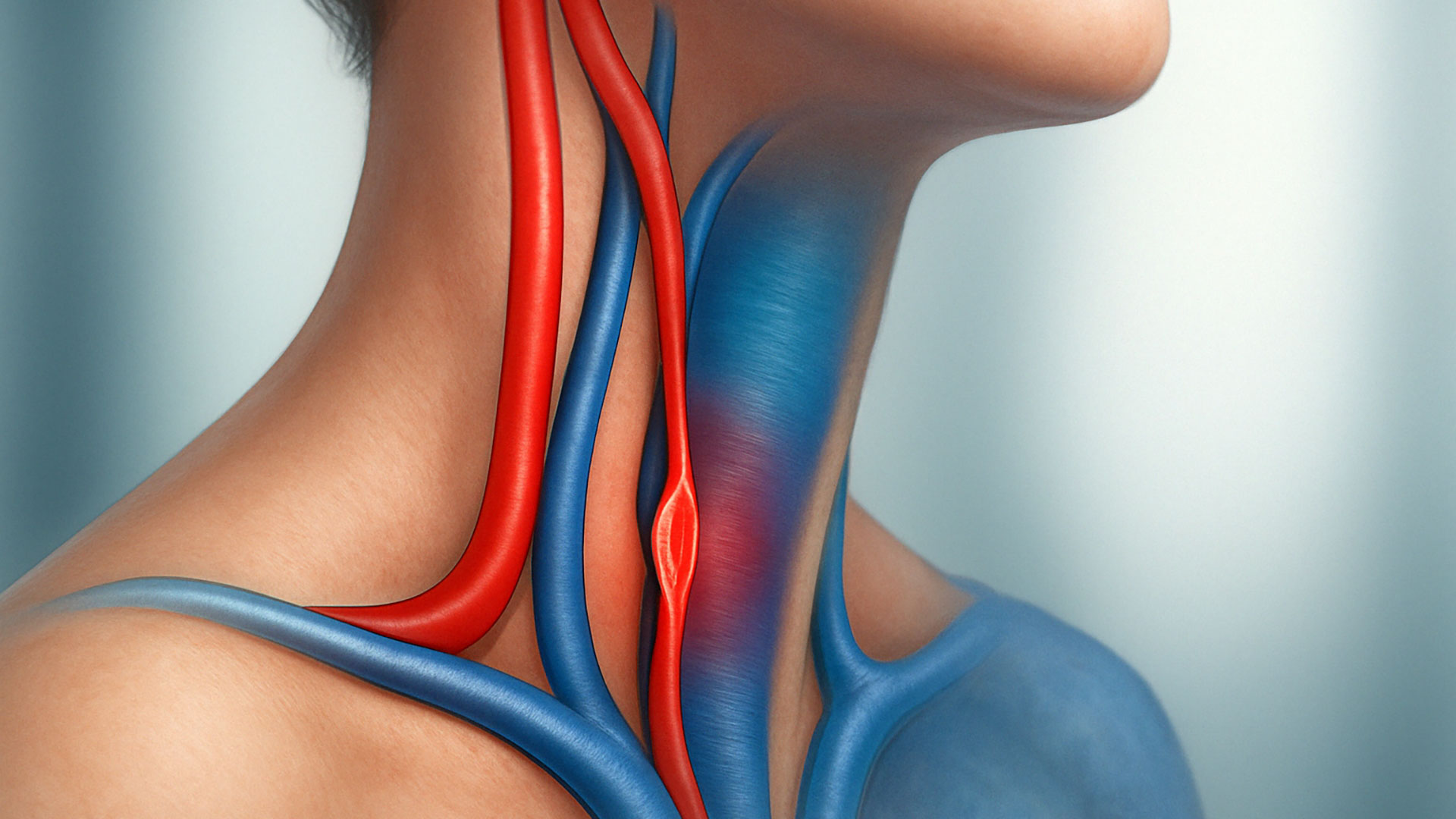
CCSVI Diagnosis: Techniques, Approaches, and a Comparison of Methods
An accurate and timely diagnosis of Chronic Cerebrospinal Venous Insufficiency (CCSVI) is a crucial step toward developing an effective, personalized treatment plan. Since CCSVI involves obstructions to venous outflow from the brain to the heart, its detection requires precise diagnostic techniques performed by experienced specialists in a dedicated clinical setting.
On this page, we’ll take a closer look at the main techniques used to diagnose CCSVI, exploring their advantages, limitations, and role in guiding the patient’s treatment pathway.
The Importance of an Accurate Diagnosis
CCSVI can present with non-specific symptoms such as persistent headaches, brain fog, dizziness, chronic fatigue, and visual or neurological disturbances. However, because these symptoms often overlap with other conditions, a specific and thorough instrumental evaluation is essential for an accurate diagnosis.
An imprecise or incomplete diagnosis can:
- portare a trattamenti inefficaci o non indicati,
- ritardare la cura di eventuali patologie associate (come la sclerosi multipla),
- trascurare compressioni venose acquisite o ostacoli interni al lume vasale.
Transcranial and Extracranial EcoColorDoppler
EcoColorDoppler is a non-invasive ultrasound examination that allows for the assessment of venous blood flow in the internal jugular veins, vertebral veins, and intracranial veins through temporal windows.
How It’s Performed
- The exam is carried out in both the supine (0°) and seated (90°) positions to observe venous dynamics under different postural conditions.
- Includes respiratory maneuvers (inhalation/exhalation) and gentle cervical compression to detect abnormal reflux.
Advantages
- Non-invasive, painless, and easily repeatable.
- Provides real-time dynamic assessment.
- Measures flow velocity, direction, and the presence of reflux or stenosis.
Limitations
- Requires a high level of technical skill from the operator.
- In some anatomies, it may be difficult to visualize deep or intracranial veins.
- It doesn’t always allow for precise identification of the cause of the obstruction (e.g., stiff valve versus external compression).
Magnetic Resonance Venography (MRV)
MRV (Magnetic Resonance Venography) is an MRI technique using contrast agents that provides a detailed mapping of the cerebral and cervical venous system.
How It’s Performed
- The contrast agent highlights the venous structures.
- Three-dimensional imaging allows for the visualization of anomalies, hypoplasia, or occlusions.
Advantages
- Provides a detailed and comprehensive view of the intracranial and extracranial venous system.
- Non-invasive, although it does require the administration of a contrast agent.
- Useful for the morphological assessment of veins.
Limitations
- It does not provide real-time dynamic information about venous flow.
- It may not detect reflux or incompetent valves.
- It's a more expensive and less widely accessible technique compared to EcoColorDoppler.
Catheter Venography (Selective Venous Catheterization)
An invasive technique considered the gold standard for diagnosing CCSVI in complex cases. It involves inserting a catheter into the venous system to inject contrast medium directly into the internal jugular and brachiocephalic veins.
How It’s Performed
- Performed in an angiography suite under local anesthesia.
- The catheter is usually inserted through the femoral vein and guided to the target veins.
Advantages
- Allows direct visualization of obstructions and their behavior during breathing and postural changes.
- It can be combined with venous angioplasty (PTA) if indicated.
Limitations
- Invasive, with a small risk of complications (hematomas, infections, contrast reactions).
- Requires a specialized interventional radiology team.
Intravascular Ultrasound (IVUS)
One of the most innovative and detailed techniques, IVUS (Intravascular Ultrasound) involves inserting a micro-ultrasound probe inside the vein, providing images from the inside out.
What It’s Used For
- Identifies stiff valves, membranes, septa, and fibrous rings.
- Distinguishes between endoluminal causes (intrinsic to the vein) and extrinsic compressions.
Advantages
- Maximum anatomical and functional precision.
- Excellent for planning endovascular treatments.
Limitations
- It's an invasive technique, typically performed alongside catheter venography.
- Available only in highly specialized centers.
Comparison of Diagnostic Methods
| Method | Invasiveness | Main Advantages | Main Limitations |
|---|---|---|---|
| EcoColorDoppler | Non-invasive | Dynamic, widely accessible | Operator-dependent |
| MRV | Non-invasive | Comprehensive view of the venous system | Static; less effective for detecting reflux |
| Catheter Venography | Invasive | High precision, allows for therapeutic intervention | Requires an angiography suite |
| Intravascular Ultrasound (IVUS) | Invasive | Detailed intraluminal imaging, visualization of valves and septa | Costly; available only in specialized centers |
Every patient with suspected CCSVI deserves a tailored diagnostic approach, starting with non-invasive methods and advancing, if necessary, to more specialized techniques. The choice will depend on the clinical picture, the symptoms, and the availability of diagnostic technologies.
In my practice, I provide a comprehensive and personalized diagnosis, combining imaging studies, clinical evaluation, and biomechanical assessment to offer patients the best possible therapeutic pathway.
Dr. Raffaello Pagani
Specialist in Vascular Surgery
📞 For a comprehensive evaluation, contact the office and schedule your appointment.