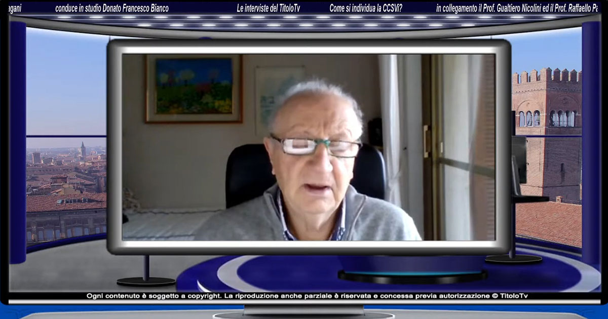
In a recent scientific exploration conducted alongside Professor Gualtiero Nicolini, Dr. Raffaello Pagani delved into the intricate relationship between CCSVI (Chronic Cerebrospinal Venous Insufficiency) and Multiple Sclerosis, providing a comprehensive and up-to-date overview of diagnosis and potential treatment strategies. The interview, designed for a broad audience, sheds light on how the traditional approach has now been surpassed and underscores the importance of a more extensive, multidisciplinary perspective on the disease.
CCSVI and MS: Looking Beyond the Valves
The concept of CCSVI, introduced in 2009 by Professor Zamboni, brought to light a new pathogenic hypothesis for multiple sclerosis: venous congestion and inadequate blood drainage from the brain to the heart could contribute to chronic inflammation and neurological damage. Initially, the focus was on malformed valves in the internal jugular vein, located in the lower neck region (J1), as the primary cause of impaired blood outflow.
However, in the years that followed, clinical experience and the use of more advanced diagnostic techniques revealed a more complex reality: venous obstructions can also occur upstream of the valves, in the middle and upper neck regions. These blockages may result from muscular compression, bone structures, adjacent arteries, or anatomical abnormalities.
Beyond Angioplasty: The Failure of a One-Sided Approach
Many patients treated in the early years with angioplasty (PTA) to dilate the venous valves experienced recurrences or unsuccessful outcomes. According to Dr. Pagani, this was due to an overly narrow understanding of the condition: focusing solely on the valves while overlooking other potential compression points—such as those caused by the omohyoid muscle or the styloid process of the temporal bone—meant missing the true underlying cause of the obstruction.
This is where the need for comprehensive diagnostics becomes clear. Beyond cervical Color Doppler Ultrasound, modern approaches now include Magnetic Resonance Angiography (MRA), CT Angiography, and venography. Crucially, the analysis must extend to the intracranial tract and the craniocervical junction (C1-C3)—areas where some of the most significant compression causes are often found.
A New Team of Specialists
To effectively address CCSVI in patients with multiple sclerosis, relying solely on vascular surgeons or angiologists is no longer sufficient. A multidisciplinary approach is now essential, incorporating specialists such as:
- Neuro-radiologists to assess intracranial venous morphology and detect signs of venous hypertension.
- Interventional radiologists, who can access deep-seated areas via catheterization—avoiding invasive surgery altogether.
- Physical medicine specialists (physiatrists) and physiotherapists, who provide conservative therapies—particularly crucial when addressing muscular compression.
- Neurosurgeons or ENT specialists, for cases requiring surgical removal of compressive bone structures—such as styloidectomy or C1-level interventions.
The evidence is clear: CCSVI is no longer viewed as a simple, isolated vascular condition—but rather a complex syndrome demanding coordinated multidisciplinary care.
The Contribution of Ophthalmologists and ENT Specialists
A key insight from the interview highlights CCSVI’s ocular and auditory manifestations. Unexplained visual disturbances—including blurred vision, papilledema, and retinal hemorrhages—or symptoms like tinnitus and vertigo may serve as indirect markers of venous obstruction.
Dr. Pagani emphasizes the critical role of ophthalmologists in CCSVI evaluation. Through fundus examination, they can detect signs of venous hypertension or retrograde blood flow. Additionally, Color Doppler Ultrasound of the optic nerve and its sheath provides valuable hemodynamic insights.
Similarly, ENT specialists play a key role when Menière’s syndrome or vestibular disorders are present – conditions where research has demonstrated clear correlations with CCSVI.
A Tailored Approach: From Diagnosis to Surgery
The key takeaway from this interview is clear: diagnosis and treatment must be tailored to each individual patient. CCSVI manifests differently in every case, and therapeutic approaches should target the primary obstruction—whether venous, muscular, or anatomical:
- Conservative therapies for muscle compressions (targeted physiotherapy, postural exercises);
- Angioplasty or stenting for pure valvular cases;
- Decompressive surgery for bone compressions (styloidectomy, procedures on C1/C2);
- Combination of techniques for complex cases.
Telemedicine and Remote Collaboration
Dr. Pagani also highlights how building a network of professionals across different locations is now entirely possible. With telemedicine and the digital sharing of medical reports and imaging, coordinating a diagnostic and therapeutic pathway—both nationally and internationally—has become simpler and more accessible than ever before.
Dr. Pagani also highlights how building a network of professionals across different locations is now entirely possible. With telemedicine and the digital sharing of medical reports and imaging, coordinating a diagnostic and therapeutic pathway—both nationally and internationally—has become simpler and more accessible than ever before.
CCSVI offers a new perspective on many complex conditions, such as multiple sclerosis. However, addressing it effectively requires a shift in mindset: focusing solely on the valves is no longer enough.
What’s needed is a broader view, a network of specialists, and thorough, in-depth diagnostics.
The interview with Dr. Pagani makes it clear: the path to improvement lies in knowledge, collaboration, and the ability to move beyond rigid, outdated approaches. It’s a message of hope and practical progress for all patients.