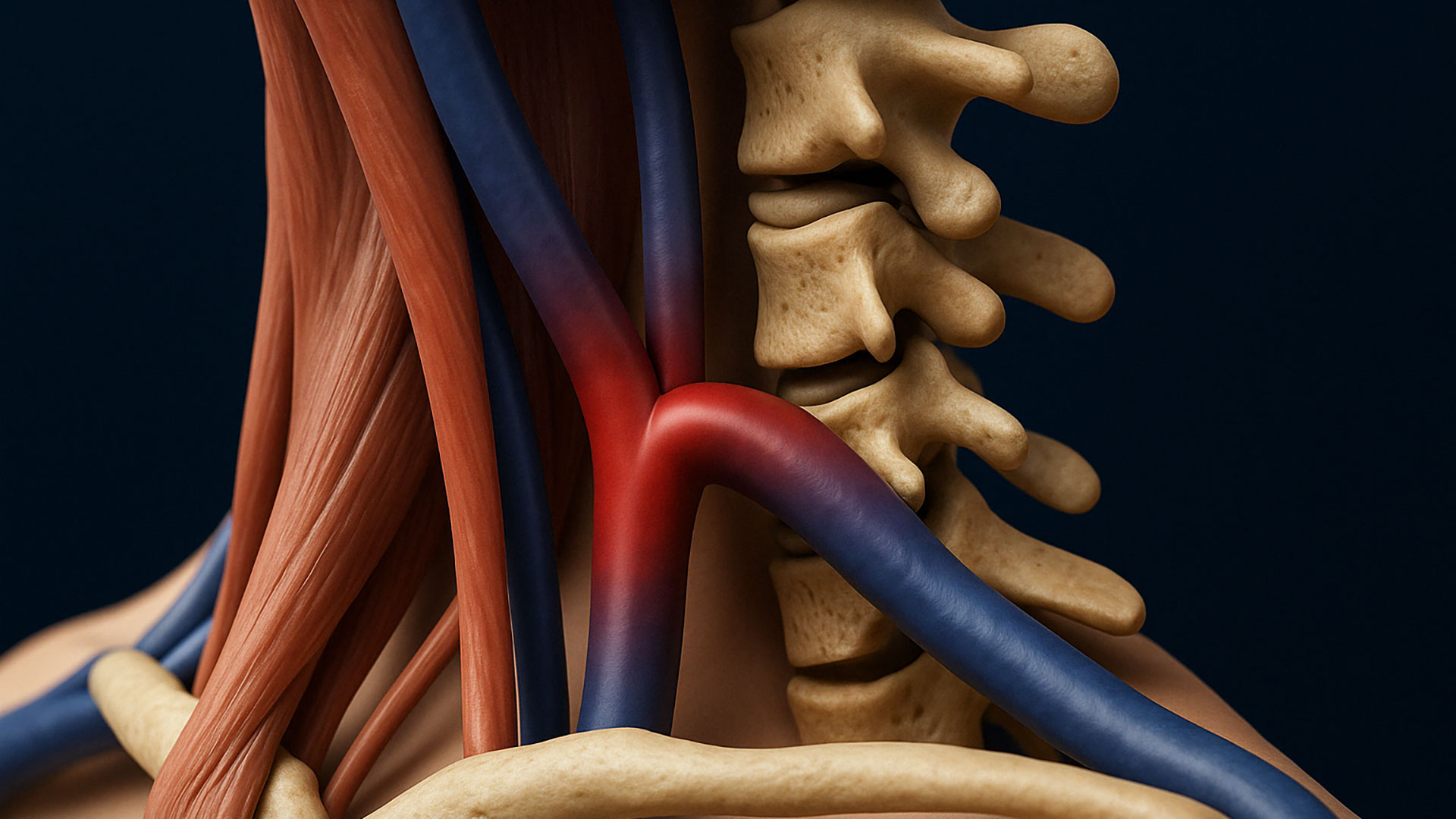
CCSVI, or Chronic Cerebrospinal Venous Insufficiency, is a vascular condition that occurs when venous blood flow from the brain to the heart is obstructed. In some patients, the issue doesn’t stem from abnormalities inside the vein itself, but rather from external pressure exerted by nearby anatomical structures. These are known as extrinsic compressions.
What Are Extrinsic Compressions?
Extrinsic compression refers to a situation where blood flow within the cerebral and cervical veins is hindered by external elements pressing against the vessel wall, narrowing its caliber or altering its flow dynamics. Unlike endogenous obstructions—such as malformed valves, septa, or membranes inside the vein—here it’s muscles, bones, or other nearby structures that apply mechanical pressure on the vein, leading to functional impairment even without any internal lesions.
The most common sites where these compressions are observed include:
- Upper cervical region, near C1 and C2
- Jugular area, at the level of the omohyoid and sternocleidomastoid muscles
- Around the styloid process of the temporal bone
- At the jugular foramen, where the veins exit the skull
In these areas, the veins—especially the internal jugular vein—can become trapped between rigid or contractile structures, leading to slowed or stagnant venous flow.
Typical Symptoms Associated with Extrinsic Compressions
Patients with CCSVI caused by external compression may experience symptoms similar to those seen in endogenous forms, but with certain dynamic features that vary depending on head or body position.
The most common symptoms include:
- Orthostatic headache, meaning head pain that worsens when standing
- Brain fog, with reduced concentration and cognitive slowing
- Paresthesias, tingling or numbness in the hands and feet
- Dizziness and imbalance, especially during neck movements
- Visual disturbances, such as blurred or double vision
- A sensation of pressure in the head or neck
These symptoms often worsen with neck movement, prolonged postures, or following cervical trauma (such as whiplash).
Diagnosing Extrinsic Compressions
An accurate diagnosis requires an integrated assessment combining vascular imaging and biomechanical analysis.
Key diagnostic tests include:
- Transcranial and extracranial EchoColorDoppler, performed in both supine and seated positions to observe posture-related changes in blood flow
- CT and MRI of the cervical spine, to identify bone abnormalities or structural deviations
- Magnetic Resonance Venography (MRV), to assess venous caliber
- Dynamic postural study, to analyze how cervical movements affect venous flow
In some cases, catheter venography or intravascular ultrasound (IVUS) can be useful to directly observe changes in the venous lumen during dynamic maneuvers.
Common Causes of Venous Compression
In the context of CCSVI caused by extrinsic compression, several anatomical structures can interfere with normal venous outflow by exerting external mechanical pressure on the major veins. Among them, the omohyoid muscle plays a significant role, as it crosses the cervical region horizontally and can compress the internal jugular vein at its midsection, known as the J2 segment. Another muscle often involved is the sternocleidomastoid, whose position and tone can affect the deeper and upper segments of the same vein.
The upper cervical vertebrae, particularly C1 and C2, are also frequently responsible for bony compressions. These become especially significant in individuals with cranio-cervical instability, a condition that can increase venous pressure in the upper drainage pathways. Additionally, an enlarged or misaligned styloid process of the temporal bone can compress the veins exiting the skull, disrupting normal blood flow.
Finally, adjacent arteries such as the internal carotid cannot be overlooked, as in some cases their positioning can directly interfere with the venous lumen, particularly in the upper segments of the neck. Although these compressions are external, they can have a significant clinical impact, especially when combined with other predisposing anatomical factors.
Treatment Options and Therapeutic Approaches
The treatment of CCSVI caused by extrinsic compression always requires an individualized approach, tailored to the specific cause of the obstruction. In cases where the compression results from muscular tension, poor posture, or joint misalignments, conservative therapies are typically the first line of intervention.
Among these, targeted physiotherapy focuses on relaxing the muscle groups involved in venous compression—such as the omohyoid and sternocleidomastoid—through stretching exercises, mobilization, and postural strengthening. Cranio-cervical osteopathy is also a valuable tool, helping to improve joint mobility and restore biomechanical balance in the cervical and occipital regions. Postural therapy plays a key role as well, assisting patients in correcting long-standing poor posture habits that can contribute to vascular compression over time.
In more complex cases, or when conservative treatment fails, more invasive interventions may be considered. Decompressive surgery is recommended when there is clear evidence of compression from bony structures, such as an enlarged styloid process or displacement of the upper cervical vertebrae. If an internal venous stenosis is also present, PTA—percutaneous transluminal angioplasty—may be considered to help restore the vein’s normal caliber. In selected situations, surgical removal of hypertrophic muscles or fibrous bands that interfere with proper venous outflow may also be an option.
The choice of treatment is always made within a multidisciplinary framework, involving various specialists such as the vascular surgeon, physiotherapist, osteopath, neurosurgeon, and, when needed, an interventional radiologist.
Structures Involved in Extrinsic Compressions
| Structure | Type | Compression Mechanism | Possible Interventions |
|---|---|---|---|
| Omohyoid Muscle | Muscular | Compression of the internal jugular vein at the mid-level (J2) | Physiotherapy, myofascial release |
| Sternocleidomastoid Muscle | Muscular | Dynamic interference with venous flow in deeper segments | Osteopathy, postural physiotherapy |
| C1 and C2 Vertebrae | Bony | Cranio-cervical instability and narrowing of the venous lumen | Decompressive surgery, postural therapy |
| Styloid Process | Bony | Extrinsic compression of the upper segments of the internal jugular vein | Surgical removal (styloidectomy) |
| Internal Carotid Artery | Vascular | Pulsation and pressure on the adjacent vein | Integrated approach, surgery in selected cases |
Extrinsic compressions are one of the most overlooked yet significant causes of CCSVI. This is especially true in younger patients, athletes, and individuals with postural abnormalities or chronic cervical dysfunction. Early identification of these compressions can prevent symptom progression and allow for the initiation of targeted, effective treatment.
A comprehensive and dynamic diagnosis is essential for distinguishing between congenital CCSVI and forms caused by external compression. Only a thorough, personalized evaluation can accurately identify the source of the venous outflow obstruction and guide the development of the most appropriate treatment plan, whether conservative or surgical.
📞 Looking for a comprehensive evaluation of venous compressions?
If you suspect that your symptoms may be linked to CCSVI caused by extrinsic compression, schedule a specialist consultation with Dr. Raffaello Pagani. With a personalized, multidisciplinary approach, you’ll receive an accurate diagnosis and a tailored treatment plan designed specifically for your case.
👉 Contact the office for more information or to schedule an appointment.